
These Stem Cells Prevent Osteoarthritis
Columbia researchers have discovered stem cells in adult mice that are essential in maintaining healthy cartilage in the joints and cause osteoarthritis when age or injury kills the cells.
The findings raise the possibility that therapies can be developed to prevent osteoarthritis by keeping the cells alive or by introducing replacement cells into the joint.
“Our study also reframes osteoarthritis as a disease of stem cell imbalance, as opposed to a strictly mechanical wear and tear disease, and opens a completely new line of investigation,” says Siddhartha Mukherjee, MD, DPhil, associate professor of medicine at Columbia University Vagelos College of Physicians and Surgeons, who co-led the study.
“Our hypothesis is that yes, of course, there's wear and tear. But that wear and tear kills the stem cells that are really preventing damage to the joint.”
Gremlin cells create and maintain cartilage
Osteoarthritis is a common degenerative disease of the joints that affects nearly 33 million American adults. The disease occurs when articular cartilage that coats and protects the bones within a joint breaks down. The loss of cartilage causes bones to grind against each other, and the underlying bone begins to change. No therapy can stop or slow the degeneration.
Mukherjee had previously discovered that certain stem cells, marked by a protein called gremlin-1, give rise to articular cartilage in the joints of mice during development. These cells were still present in a thin layer in the joint as the animals matured, and Mukherjee wondered if they had any role in osteoarthritis.
In the new study, together with a team of Australian collaborators, Mukherjee and Toghrul Jafarov, PhD, an associate research scientist in the lab, showed that these gremlin stem cells are essential to the health of the cartilage as the mouse matures. After genetically ablating the gremlin cells in the articular cartilage of mature mice, the researchers found that the cartilage degenerated and the animals developed severe arthritis.
Gremlin cells were also lost from the joint after injury, which is known to trigger osteoarthritis, and as the animals aged.
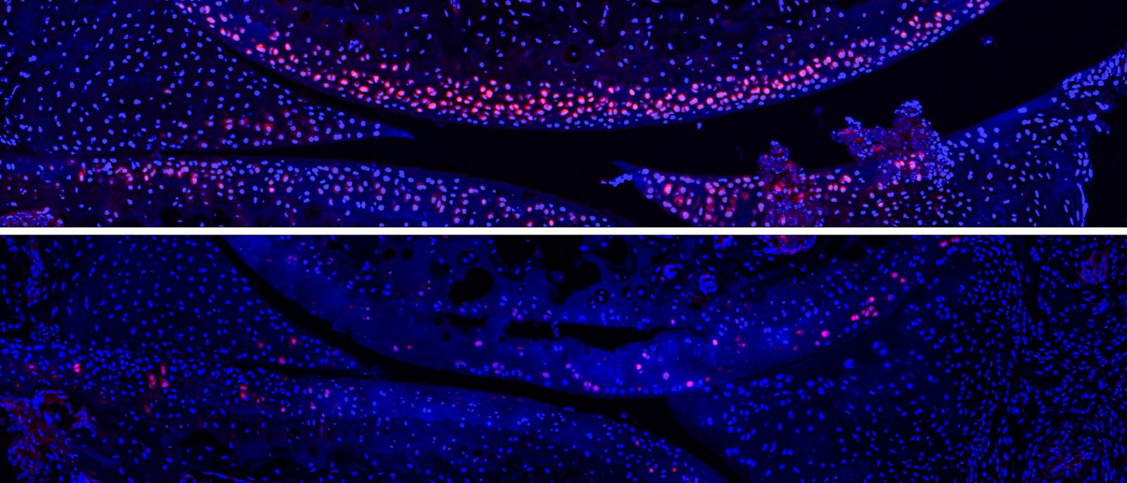
Gremlin progenitor cells (stained pink) prevent osteoarthritis. In the upper panel, the gremlin progenitor cells line the outer edges of bones that make up a knee joint. In the lower panel, the cells have disappeared. Images courtesy of Toghrul Jafarov / Columbia University Irving Medical Center.
Rejuvenating gremlin cells prevents osteoarthritis
Even though injury and age killed the gremlin cells over time, the researchers found that the cells can be rescued with a growth factor called FGF18.
FGF18 injections increased the number of gremlin cells in the joint, led to thicker cartilage, and protected the mice from osteoarthritis.
Under the name sprifermin, FGF18 is already being tested in clinical trials although, until now, how FGF18 worked was unclear. “We’ve shown that the most likely target of FGF18 is the gremlin progenitor cell,” Mukherjee says.
Working with Abdullah Ali, PhD, assistant professor of medical sciences, Jafarov has also found additional targets in the gremlin cells that may have even greater therapeutic potential.
Next steps: What kills the gremlin cells?
Regenerative therapies may only work when gremlin cells are still present in the joint. “That’s one difficult thing about regenerative medicine: By the time a patient has symptoms, there’s nothing left to save. The things we could act on are dead,” Mukherjee says.
“You have to find that sweet spot, when cells are still alive,” Jafarov adds. “So it’s important for us to pinpoint when the gremlin cells become impaired and what is killing these cells, particularly during aging.”
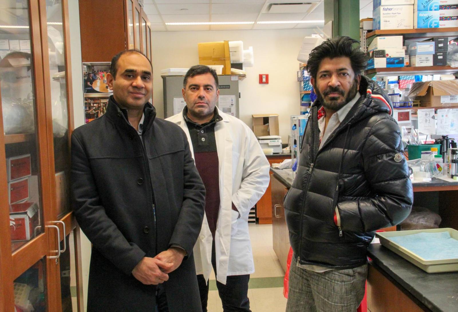
Columbia researchers Abdullah Ali, left, Toghrul Jafarov, center, and Siddhartha Mukherjee are trying to identify when gremlin cells disappear from joints and how the cells could be protected or implanted to prevent osteoarthritis. Photo from Columbia University Irving Medical Center.
One theory is that inflammation kills the cells, another is that the cells have an innate regenerative capacity and become dormant after they hit their limit. “If we can learn how and when the cells begin to die, we may be able to intervene then to keep them active for longer,” Jafarov says.
The researchers are also beginning to investigate ways to implant gremlin cell into joints affected by osteoarthritis.
Tissue engineers have had high hopes for repairing damaged cartilage with stem cell implants, but so far these attempts have been unsuccessful, possibly because engineers have selected the wrong stem cells.
“Identifying the right cell to target or use in cell therapy is critical,” Mukherjee says. “If you’re looking at the wrong cells, then you’re not identifying the important pathways that can lead to a treatment.”
References
More information
All authors: Jia Q. Ng (University of Adelaide), Toghrul H. Jafarov (Columbia), Christopher B. Little (University of Sydney), Tongtong Wang (University of Adelaide and South Australian Health and Medical Research Institute), Abdullah Ali (Columbia), Yan Ma (Columbia), Georgette A Radford (University of Adelaide), Laura Vrbanac (University of Adelaide), Mari Ichinose (University of Adelaide), Samuel Whittle (University of Adelaide), David Hunter (University of Sydney), Tamsin RM Lannagan (University of Adelaide), Nobumi Suzuki (University of Adelaide), Jarrad M. Goyne (South Australian Health and Medical Research Institute), Hiroki Kobayashi (University of Adelaide), Timothy C. Wang (Columbia), David Haynes (University of Adelaide), Danijela Menicanin (University of Adelaide), Stan Gronthos (University of Adelaide), Daniel L. Worthley (South Australian Health and Medical Research Institute), Susan L. Woods (University of Adelaide and South Australian Health and Medical Research Institute), and Siddhartha Mukherjee (Columbia).
This research was funded in part through grants from the U.S. National Institutes of Health (Cancer Center Support Grant P30CA013696, the National Center for Advancing Translational Sciences grant UL1TR001873, and R01AR069852), the National Health and Medical Research Council of Australia (APP1099283), Cancer Council SA Beat Cancer Project, and the State Government of South Australia (MCF0418); and an Endeavour Research Fellowship from the Australian government (ERF_RDDH_179965).
The authors declare no competing interests.