Study Points Toward New Ways to Prevent Liver Cancer
Almost all liver cancers develop after decades of chronic liver disease, but a new discovery by Columbia researchers may lead to treatments that could break the link.
The new research shows that during chronic liver disease a shift in the balance of quiescent and activated stellate liver cells not only promotes fibrosis but also sets the stage for the most common type of primary liver cancer, called hepatocellular carcinoma.
The findings, published online in Nature, suggest that it may be possible to prevent the development of liver cancer—the fourth-leading cause of cancer death worldwide—by interfering with stellate cell activation.
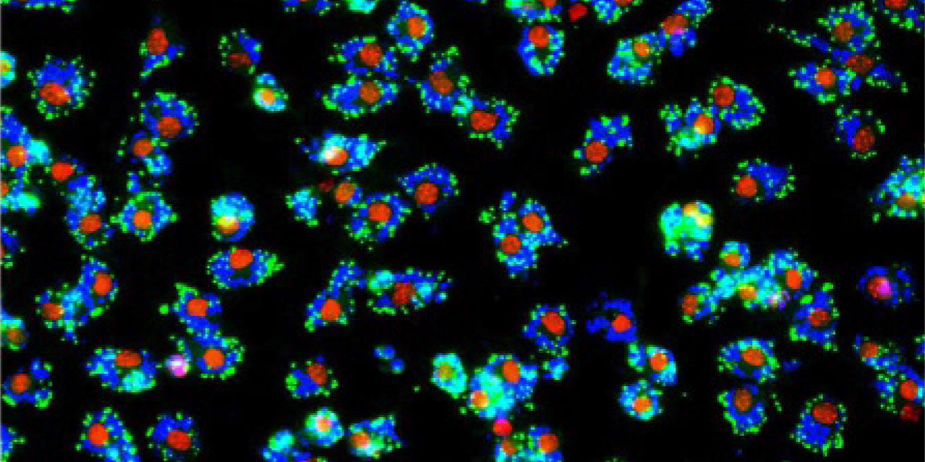
Stellate liver cells have two identities: protective and tumor-promoting. During chronic liver disease, a dynamic shift in the balance of cells toward tumor-promoting cells sets the stage for liver cancer. Image of stellate cells from Robert Schwabe lab.
Hepatocellular carcinoma develops almost exclusively in patients with a chronic liver disease, including cirrhosis, hepatitis, or non-alcoholic fatty liver disease. These diseases often cause extensive and progressive scar tissue (aka fibrosis) in the liver.
“Although fibrosis almost always precedes the cancer, little is known about causal relationships between the two and how we might be able to interrupt this process,” says study leader Robert F. Schwabe, MD, professor of medicine and director of the Digestive and Liver Disease Research Center at Columbia University Vagelos College of Physicians and Surgeons.
Schwabe and his team focused on the liver’s stellate cells, so named for the multiple protrusions that give them a star-like shape, because they are heavily involved in liver fibrosis.
After developing new genetic tools that allow researchers to manipulate and analyze stellate cells in mice, Schwabe discovered that the population of these cells changes as the cancer develops.
“In a healthy liver, stellate cells are quiescent and express factors, notably hepatocyte growth factor, that protect the organ,” he says. “In the presence of chronic liver disease, however, the number of the protective quiescent stellate cells decreases while more stellate cells become activated.”
Activated cells express factors that promote tumors, particularly collagen, the building block of scar tissue. This dynamic shift from protective to tumor-promoting cells sets the stage for liver cancer.
The same imbalance in stellate cells and its association with increased risk for cancer development was observed in patients with liver cancer, suggesting the same process occurs in people.
Hepatocellular carcinomas are typically detected too late to be cured, and most patients die within two years of diagnosis despite new, more efficient medical therapies such as the combination of atezolizumab and bevacizumab. The incidence of hepatocellular carcinoma has tripled in the past 30 years and is expected to increase further due to rising rates of non-alcoholic fatty liver disease that is driven by rising obesity.
The discovery raises the prospect of preventing liver cancer by restoring the normal balance of stellate cells.
“I don’t think we will be able to treat cancer by restoring stellate cell balance,” says Schwabe. “Activated stellate cells create an environment that is favorable for cancer development, but they do not seem to play a role in cancer progression once the tumor has developed.
“But restoring the balance of cells or associated mediators in people with chronic liver disease may reduce the risk of tumors developing in the first place. This would be a huge benefit for the many millions of people with chronic liver disease, who are highly likely to develop liver cancer.”
References
The study is titled “Opposing roles of hepatic stellate cell subpopulations in hepatocarcinogenesis” and was published in Nature on Oct. 5.
All authors: Aveline Filliol (Columbia), Yoshinobu Saito (Columbia), Ajay Nair (Columbia), Dianne H. Dapito (Columbia), Le-Xing Yu (Columbia), Aashreya Ravichandra (Columbia), Sonakshi Bhattacharjee (Columbia), Silvia Affo (Columbia), Naoto Fujiwara (University of Texas Southwestern Medical Center), Hua Su (University of California, San Diego), Qiuyan Sun (Columbia), Thomas M. Savage (Columbia), John R. Wilson-Kanamori (University of Edinburgh), Jorge M. Caviglia (Columbia), LiKang Chin (University of Pennsylvania), Dongning Chen (University of Pennsylvania), Xiaobo Wang (Columbia), Stefano Caruso (Sorbonne Université), Jin Ku Kang (Columbia), Amit Dipak Amin (Columbia), Sebastian Wallace (University of Edinburgh), Ross Dobie (University of Edinburgh), Deqi Yin (Columbia), Oscar M. Rodriguez-Fiallos (Columbia), Chuan Yin (Columbia), Adam Mehal (Columbia), Benjamin Izar (Columbia), Richard A. Friedman (Columbia), Rebecca G. Wells (University of Pennsylvania), Utpal B. Pajvani (Columbia), Yujin Hoshida (University of Texas Southwestern Medical Center), Helen E. Remotti (Columbia), Nicholas Arpaia (Columbia), Jessica Zucman-Rossi (Sorbonne Université), Michael Karin (University of California, San Diego), Neil C. Henderson (University of Edinburgh), Ira Tabas (Columbia), and Robert F. Schwabe (Columbia).
This work was supported by the NIH (grants R01CA190844, R01CA228483, R01DK116620, 1P30DK132710, CA233794, RR180016, R37CA258829, R21CA263381, F31 DK091980); a Deutsche Forschungsgemeinschaft grant (GZ:BH 155/1-1); the Ligue Nationale contre le Cancer (Equipe Labellisée) and Labex OncoImmunology (Investissement d’avenir); a Wellcome Trust Senior Research Fellowship in Clinical Science (ref. 219542/Z/19/Z); the Medical Research Council; a Chan Zuckerberg Initiative Seed Network Grant; a Foundation pour la Recherche Medicale postdoctoral fellowship (SPE20170336778); an American Liver Foundation Postdoctoral Research Award; an International Liver Cancer Association Fellowship; a Mandl Connective Tissue Research Fellowship; the Uehara Memorial Foundation; the Russell Berrie Foundation; an American Liver Foundation Postdoctoral Research Fellowship Award; a Cholangiocarcinoma Foundation Innovation Award; and a Research Scholar Award from the American Gastroenterological Association. These studies used the resources of the Herbert Irving Comprehensive Cancer Center funded in part through NIH grants P30CA013696 and S10OD020056.