
Individual Receptors Caught in the Act of Coupling
A new imaging technique developed by scientists at Columbia University Vagelos College of Physicians and Surgeons and St. Jude Children’s Research Hospital captures movies of receptors on the surface of living cells in unprecedented detail and could pave the way to a trove of new drugs.
The researchers used the technique to zoom in on individual receptor proteins on the surface of living cells to determine if the receptors work solo or come together to work as pairs. This work appeared in the April issue of Nature Methods.
“If two different receptors come together to form a dimer with distinctive function and pharmacology, this might allow for a new generation of drugs with greater specificity and reduced side effects,” says Jonathan Javitch, MD, PhD, the Lieber Professor of Experimental Therapeutics in Psychiatry at VP&S.
G-protein coupled receptors (GPCRs) are some of medicine’s most important molecules: About one-third of today’s drugs work by targeting a GPCR. The possibility that GPCRs form heterodimers—consisting of two different flavors of GPCR—is an especially exciting prospect for the development of better drugs.
“The potential of GPCR heterodimers for improved pharmacotherapies, including for disorders such as schizophrenia and depression, is exciting and has drawn us to the field,” Javitch says.
But for decades, scientists have hotly debated whether most GPCRs form dimers or work alone. Much of this impasse stemmed from the relatively poor spatial resolution of current techniques. Different GPCRs in a cell have been captured near each other, but it was unclear if the receptors were working together.
“The controversy over receptor dimerization has only grown fiercer with conflicting data from different labs using different methods,” Javitch says.
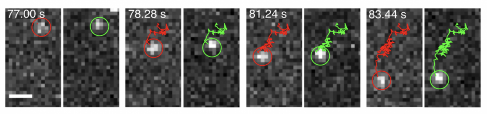
The trajectories of two paired g-protein coupled receptors on the cell surface are shown as red and green lines in the image sequence.
Using a new, more powerful technique based on single-molecule fluorescence resonance energy transfer (smFRET), Javitch and Scott C. Blanchard from St. Jude Children’s and Weill Cornell show that dimers can be tracked as they move on the cell surface and how long they last. This method takes advantage of a change in fluorescence that occurs when proteins, labeled with different fluorescent markers, are extremely close to each other. The resolution in this approach is more than 10 times greater than previous techniques.
This new and exciting technique entails multiple innovations in dyes, labeling technology, protein engineering, imaging, and software that enabled tracking of individual and coupled receptors.
Not only does this method detect GPCR dimers, it also allows, for the first time, a clear view of how receptors in a living cell change shape when activated. This will provide researchers a better understanding of how drugs can differentially impact the same receptors.
“With this method, we can now explore receptor interactions and activation mechanisms with unprecedented resolution, giving us an opportunity to investigate new therapeutic approaches,” Javitch says.
References
The research was published in a paper titled “Single-molecule FRET imaging of GPCR dimers in living cells” in Nature Methods.
All authors: Wesley B. Asher (Columbia), Peter Geggier (Columbia), Michael D. Holsey (Columbia), Grant T. Gilmore (University of Akron), Avik K. Pati (St. Jude Children’s Research Hospital), Jozsef Meszaros (Columbia), Daniel S. Terry (St. Jude Children’s), Signe Mathiasen (Columbia), Megan J. Kaliszewski (University of Akron), Mitchell D. McCauley (University of Akron), Alekhya Govindaraju (New York State Psychiatric Institute), Zhou Zhou (Weill Cornell and Queensborough Community College), Kaleeckal G. Harikumar (Mayo Clinic), Khuloud Jaqaman (University of Texas Southwestern Medical Center), Laurence J. Miller (Mayo Clinic), Adam W. Smith (University of Akron), Scott C. Blanchard (St. Jude Children’s and Weill Cornell), and Jonathan A. Javitch (Columbia).
This work was supported by the U.S. National Institutes of Health (grants MH54137, R15EY024451, R35GM119619 and 7R01GM098859-09); Hope for Depression Research Foundation; Lieber Center for Schizophrenia Research; Brain and Behavior Research Foundation NARSAD Young Investigator Award; National Science Foundation (grant CHE-1753060); UTSW Endowed Scholars Program; and the Single-Molecule Imaging Center at St. Jude Children’s Research Hospital.
Scott Blanchard has an equity interest in Lumidyne Technologies. The other authors have no competing interests.