3-D Knee
A Custom-Printed 3-D Scaffold Puts Healing on the Fast Track
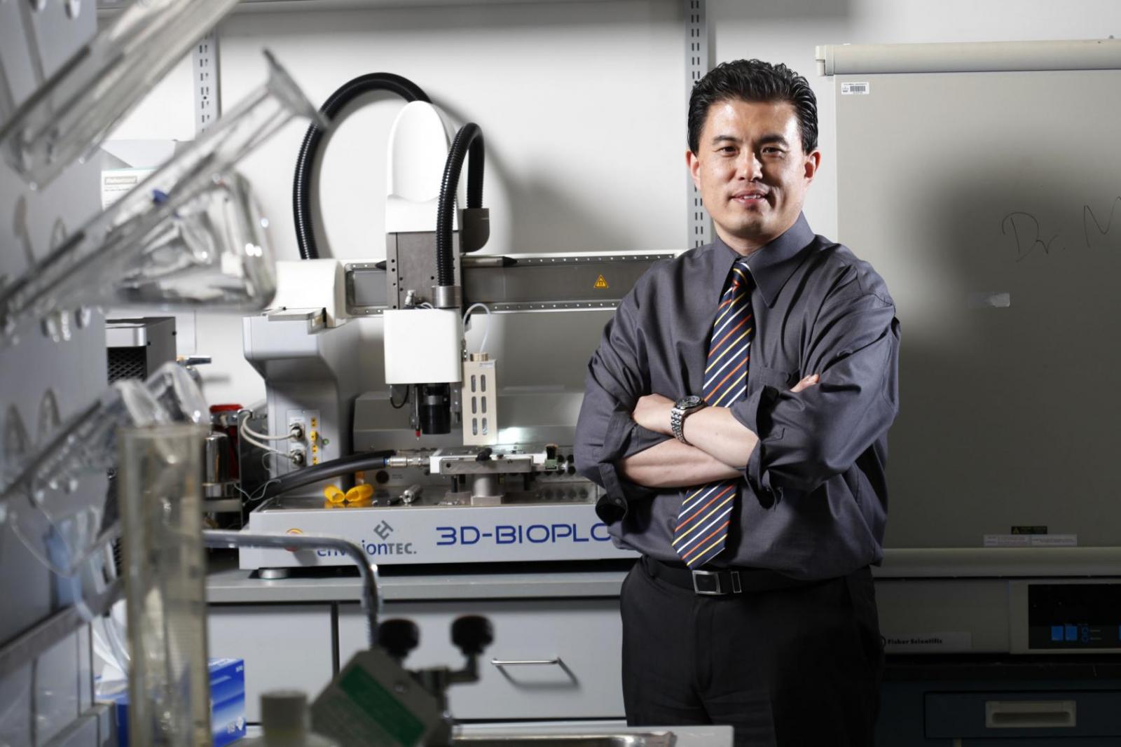
Humming on a bench in the lab of Jeremy Mao, DDS, PhD, a white robotic arm extends from a computer. This is the fourth-generation model of a 3-D printer custom made by a boutique German firm. In about the time it takes to eat dinner, the contraption can print a hydrogel in the exact shape of a knee meniscus belonging to any person—or other decent-sized mammal—at a resolution of about 20 micrometers.
Dr. Mao, professor of dentistry and of orthopedic surgery, bought the first iteration of the device back in 2007 when he came to Columbia. One of his main interests was to find a way to regenerate damaged cartilage in joints. At the time, many researchers thought that the way to regrow tissue was to transplant cells. Dr. Mao believed that prodding the body’s own stem cells to kick into gear would be an alternative. It took a few years of tinkering, but in 2010 his team reported in The Lancet that when loaded with the correct growth factors, a 3-D printed piece of biomaterial in the shape of shoulder joint cartilage could regenerate the joint in rabbits.
Next, Dr. Mao turned to the meniscus. For many people who irreparably damage this crescent of cartilage in the knee—an injury prevalent in athletes and older people—the treatment is often simply to remove the tissue, a protocol that practically guarantees that the person will develop osteoarthritis down the line. Some patients receive a meniscus transplanted from a cadaver, but such allografts are rare to come by and can never be a perfect fit. As he had with the rabbit shoulder cartilage, Dr. Mao wondered whether he could coax the body to grow its own replacement meniscus.
The meniscus is a special form of cartilage made up of cells called fibrochondrocytes. Apart from the meniscus, these cells are present only in the spine and in places where tendons connect to bone—all structures that heal poorly. The lab spent 18 months identifying the protein cues that push stem cells to differentiate into fibrochondrocytes, in a typical molecular biology experiment. Armed with that knowledge, the researchers partnered with orthopedic surgeons and veterinary scientists to make MR images of the intact meniscus in sheep and create a 3-D printed polymer copy. Then they loaded this bioscaffold with the proteins and implanted it into the sheep’s damaged knee. “In just about all the ways we look at this, the regenerated meniscuses were similar to the native ones,” Dr. Mao says. In December, Science Translational Medicine published the team’s analysis of the new technique. After three months, they reported, the sheep who received the bioscaffold implants seemed to walk just fine.
A few gaps must be bridged before testing begins in humans. Ideally, as the implanted biomaterial degrades, the stem cells generate replacement tissue, he says. “So we need to better synchronize the process of tissue degradation with new tissue formation.” The protocol also may require modification based on patient characteristics, such as age. In younger people, stem cells tend to be more abundant; for older patients, the dosage of proteins may have to be fine-tuned for aging patients. Dr. Mao hopes to start human testing in about a year once funding is in place to pursue the project.
In addition to its clinical advantages, the 3-D printing approach may represent a cost savings compared with conventional manufacture of medical devices such as metal joint implants. Despite having billion-dollar factories, conventional medical device manufacturers can take weeks or months to process an order.
Dr. Mao’s printer cost about $300,000 and was designed to be exponentially faster. “So scale-wise, it can work,” he says. “If you can print a piece of cartilage or a piece of bone, you can readily have 200 of them lined up. It’s viable.” Dr. Mao is convinced that his current work hardly even scratches the surface of the potential for 3-D printing to improve patients’ lives. “When we first got the printer, I thought, ‘Gee, this is going to be really powerful,’” says Dr. Mao. “Now more and more people are seeing the utility of 3-D printing and there’s a tremendous excitement for its use in medicine.”
Several of his colleagues at Columbia have spoken with Dr. Mao about strategies to incorporate 3-D printing into their own work. In February, he launched a collaboration with Michael Shen, PhD, professor of medicine and of genetics & development at the Herbert Irving Comprehensive Cancer Center, to print 3-D hydrogels that model the niche in which cancer cells live. In preliminary experiments, Dr. Shen found huge differences in the behavior of cancer cells in different niches. “I proposed that we could change the cancer cells’ environment,” says Dr. Mao. “If we create hundreds of different conditions in the gel, we could figure out a way to restrict cancer cell growth.”
The most obvious applications, however, lie in the field of regenerative medicine. For example, Michael Kazim, MD, clinical professor of ophthalmology and of surgery, recreated the orbital structure of the eye, an application for which the materials currently used are outdated and not biocompatible. Francis Lee, MD, the Robert E. Carroll and Jane Chace Carroll Laboratories Professor of Orthopedic Surgery, is testing simple 3-D implants to correct bone defects.
Ultimately, however, Dr. Mao predicts that the impact of 3-D printing will be even bigger—printing customized organs for transplant or printing matrices to help the body grow its own. “When that happens—and I’m convinced it will,” he says, “organ donation will become obsolete.”
https://www.youtube.com/watch?v=srVZRU6d40Y&feature=youtu.be